Imaging
OCT, OCTA, fundus photography and the technologies changing what clinicians can see.
Build the mental model first.
Start with the modality that made retinal structure measurable in cross-section.
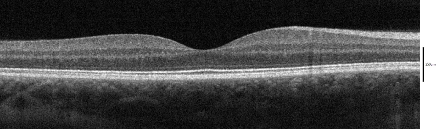
What Is OCT? A Modern Guide to Optical Coherence Tomography
How optical coherence tomography turns reflected light into cross-sectional maps of the retina—and why those maps changed retinal care.
How the major modalities differ.
Structure, flow, field of view and acquisition architecture each answer a different retinal question.
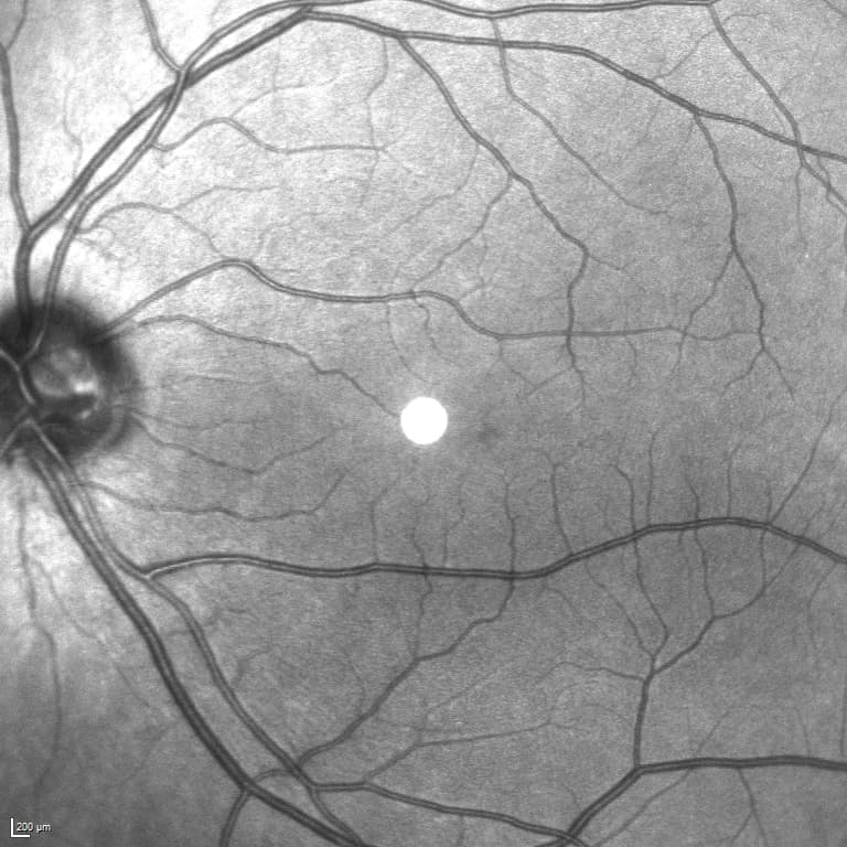
OCT vs. OCTA: What’s the Difference?
They share the same optical platform, but one primarily maps retinal structure while the other derives information about motion and blood flow.
OCT Angiography Explained: How OCTA Maps Blood Flow Without Dye
OCTA creates depth-resolved vascular maps from repeated OCT scans. The elegance is real—but so are the assumptions built into the image.
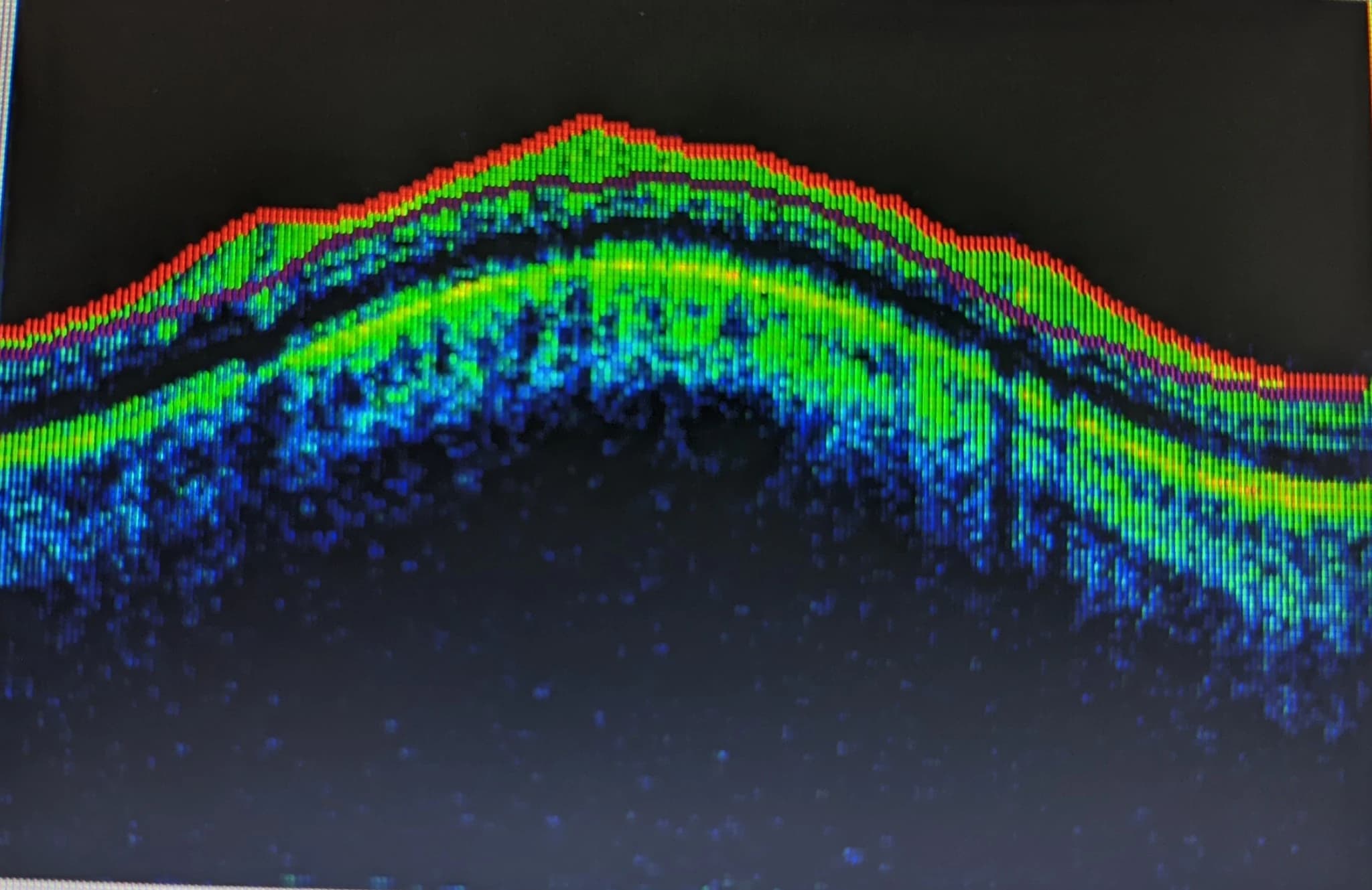
Swept-Source vs. Spectral-Domain OCT: What Actually Changes?
Both can produce excellent retinal scans. The meaningful differences are in acquisition architecture, wavelength, speed, depth and how measurements behave across devices.
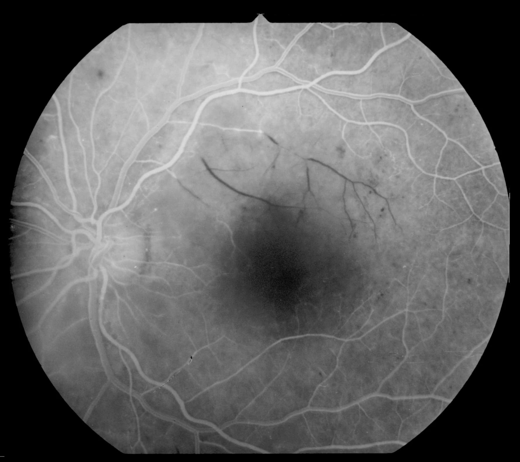
Fluorescein Angiography vs. OCTA: Why Dye Angiography Still Matters
OCTA offers depth-resolved vascular maps without an injection. Fluorescein angiography still shows something OCTA cannot: dynamic dye transit and leakage.
The scan is not self-interpreting.
Artifacts, segmentation and signal quality can change what a retinal image appears to show.
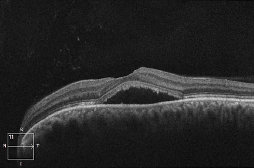
Seeing wider. Seeing smaller.
Ultra-widefield systems expand retinal context while adaptive optics pushes toward cellular-scale measurement.
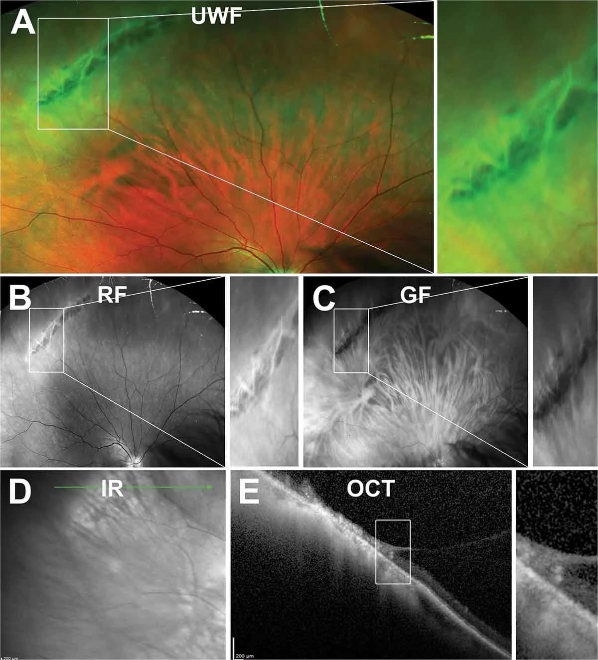
Ultra-Widefield Retinal Imaging: What We Gain by Seeing the Periphery
A larger retinal field can reveal lesions conventional photography misses. The harder question is which peripheral findings change prognosis or care.

Adaptive Optics Retinal Imaging: Seeing the Retina Cell by Cell
Adaptive optics corrects the eye’s own optical aberrations, pushing in-vivo retinal imaging toward cellular scales. The research promise is ahead of routine clinical use.
The image is evidence—not the whole answer.
Every modality is a measurement filtered through optics, acquisition and software. We explain what a scan can show, what it cannot show, and where artifacts or processing assumptions can change interpretation.